Orion™: High Dose Rate (HDR) Brachytherapy
High dose rate (HDR) brachytherapy, utilizing iridium-192 radioactive sources, is a form of radiotherapy and a standard option for the curative treatment of many forms of cancer. 10 HDR brachytherapy involves temporarily placing an applicator into the patient and then using medical imaging to identify their location in relation to the cancer and surrounding healthy tissue. A radioactive source is placed inside the applicator and irradiates the immediate vicinity of the cancer before being removed. The popularity of HDR has increased in recent years due to its effectiveness and convenience; and the majority of Radiation Oncology practices in the US are able to offer the technique.
The radioactive source is attached to the end of a wire that is housed in a radiation shielded system; during treatment, the source is automatically passed into applicators that have been pre-inserted into the patient, stopping at pre-planned locations for calculated periods of time. Once the treatment is complete the radioactive source is retracted into the shielded system and the applicators are removed from the patient.
Compared with CT, MRI is a superior imaging modality for anatomical delineation. This facilitates the improvement of radiation dose distribution for both the tumor targeted for treatment and for avoidance of normal tissue when utilizing HDR brachytherapy. 11
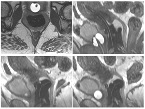
Previously the identification of an applicator’s position in the patient was achieved through MRI and CT imaging of metal wire markers that were temporarily placed in the applicator. These markers should provide two key pieces of information on an applicator’s position; the path that the radioactive source will take when placed in the applicator, and the precise location of the tip of the applicator. The exact location of this tip provides essential information from which the overall radiation dose is then calculated. While CT images provide information on the position of the metal wire marker, they are of limited value in defining the anatomy around the target tumor. MRI provides excellent anatomical image resolution but has not been able to reliably identify the metal wire marker in the applicator. Therefore, a marker was needed that provided both excellent anatomy and precise applicator positioning from a single image – this clinical need led to the development of Orion™.
Orion™
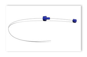
Orion™ is a novel, flexible planning marker for HDR brachytherapy that utilizes the encapsulation of the C4 positive-signal MRI agent, offering effective positive-signal MR imaging. Orion™ is intended to facilitate the identification of HDR brachytherapy applicators after placement into a patient.
Orion™ utilizes high density polyethylene (HDPE) as an encapsulating polymer. HDPE is flexible and allows for a long, thin device that can effectively replace the metal wire markers currently used when planning HDR brachytherapy. The flexible nature of Orion™ allows it to define the curved path of various applicator designs used in HDR brachytherapy.
References
- Viswanathan AN and Erickson BA. Three-dimensional imaging in gynecologic brachytherapy: A survey of the American Brachytherapy Society. Int J Radiat Oncol Biol Phys. 2010;76(1):104–109.
- Wang, MD, Ph.D., et al. A Novel Positive Contrast Magnetic Resonance Imaging Planning Marker for High Dose Rate Image Guided Brachytherapy. Semin Radiat Oncol. 2014;24(3):181–191.
© 2019 C4 Imaging.
Sirius and Orion are trademarks of C4 Imaging LLC.

